Update your subscriptions, modify your password or e-mail address, or stop subscriptions at any time on your Subscriber Preferences Page. You will need to use your e-mail address to log in. If you have questions or problems with the subscription service, please contact Subscriber Help. This service is provided to you at no charge by National Institute of Allergy and Infectious Diseases (NIAID).
|
Monday, December 21, 2020
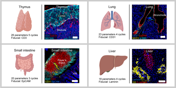
IBEX: A New Open-Source Method for Multiplex Tissue Imaging

Subscribe to:
Post Comments (Atom)
No comments:
Post a Comment